KNEE · LIGAMENT & MENISCUS SURGERY
ACL reconstruction with meniscus repair
A torn ACL and a torn meniscus in the same knee is the commonest combined injury we treat. Whether the meniscus can be repaired, and how that changes the operation and the recovery, is one of the most important decisions in knee surgery.
Dr. Prashant Parate · Shoulder, Knee & Sports Injury Surgeon · Jupiter Hospital (Baner) & Sainath Hospital (Moshi, PCMC)
Why the two injuries happen together
The ACL and the meniscus work together. The ACL prevents the shin bone from sliding forward. When it tears, the knee moves abnormally, and the meniscus takes loads it was not designed for. In the acute injury, the same twist that tears the ACL often tears the meniscus at the same time. In a chronic ACL-deficient knee, the meniscus slowly wears from the abnormal movement, and a second giving-way episode can tear it.
Roughly half of all ACL injuries have a meniscus tear on MRI, and that proportion rises the longer the ACL remains unrepaired. This is one of the reasons delayed ACL reconstruction in a young, active patient carries real risk: with repeated instability and increasing time from injury, a previously repairable meniscus can sustain further tearing and become more difficult, or sometimes impossible, to preserve.
Why repairing the meniscus matters
The meniscus is a shock absorber. Each knee has two: the medial meniscus on the inner side and the lateral meniscus on the outer. Together they spread the load across the joint surface, protect the cartilage, and contribute to knee stability.
Remove the meniscus and the load on the underlying cartilage increases dramatically. Over time that leads to arthritis, and it happens faster in a younger patient because they have more years of loading ahead. Every meaningful amount of functioning meniscus preserved today may contribute to better cartilage protection over the years ahead.
The lateral meniscus deserves particular mention. It is especially important in an ACL-injured or reconstructed knee because of its role in rotational stability. Losing the lateral meniscus in a young patient with an ACL reconstruction changes the long-term outlook of that knee significantly.
This is the principle behind our approach: repair first, and excise later only what is genuinely not repairable. Even when the chances of healing are uncertain, we will attempt a repair if there is any reasonable prospect, because the cost of a failed repair that then requires trimming is far less than the cost of an unnecessary meniscectomy that accelerates arthritis.
Blood supply determines healing
The meniscus is not equally alive throughout. Blood supply enters from the outer rim and penetrates inward, creating three zones that determine whether a tear can heal after repair.
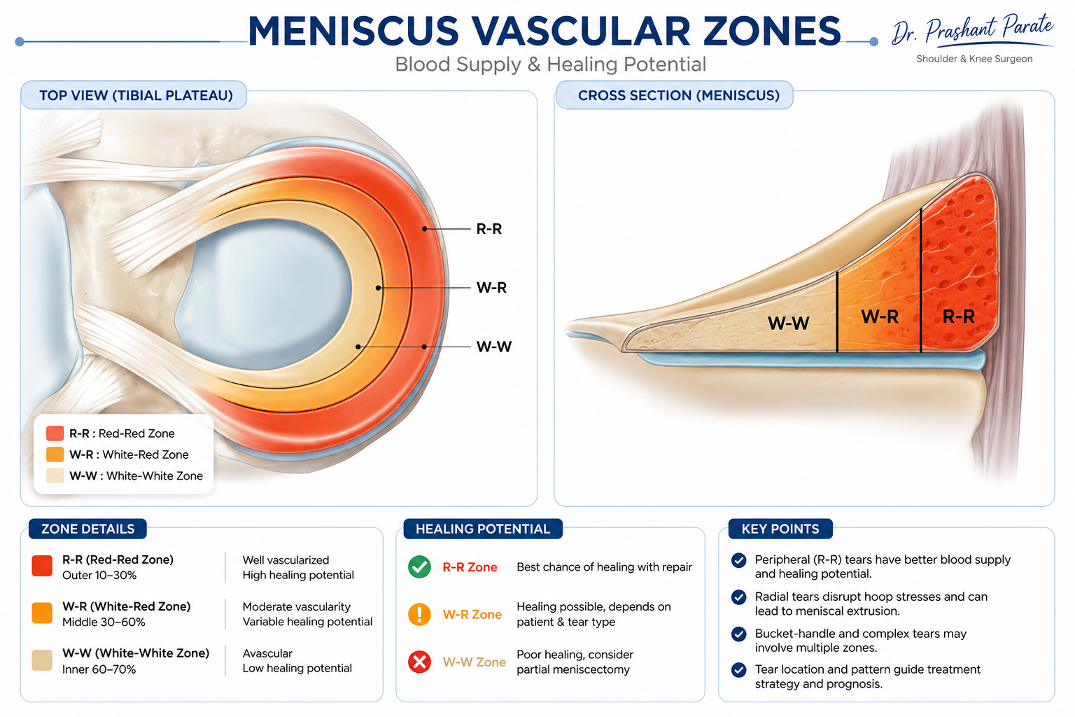
Tears in the red-red zone, the outer rim, have good blood supply and heal well after repair. Tears in the red-white zone have some blood supply and are worth repairing, particularly in younger patients and when the ACL is being reconstructed at the same time, because the surgery itself brings blood and growth factors into the joint. Tears in the white-white zone have very limited blood supply and therefore lower biological healing potential. Traditionally many were excised, although selected tears may still be considered for repair depending on the pattern, tissue quality, patient age and associated ACL reconstruction.
ACL reconstruction actually helps meniscus healing. The drilling, the bleeding and the biological response to surgery create an environment that is more favourable for repair than the resting knee. This is one reason to repair the meniscus at the time of ACL reconstruction rather than staging the procedures. The choice of graft is another important part of ACL surgery. Read more about ACL graft selection and how the right graft is chosen.
Tear patterns: which can be repaired and which cannot
The pattern of the tear matters as much as its location. Some patterns hold sutures well and heal reliably. Others do not.
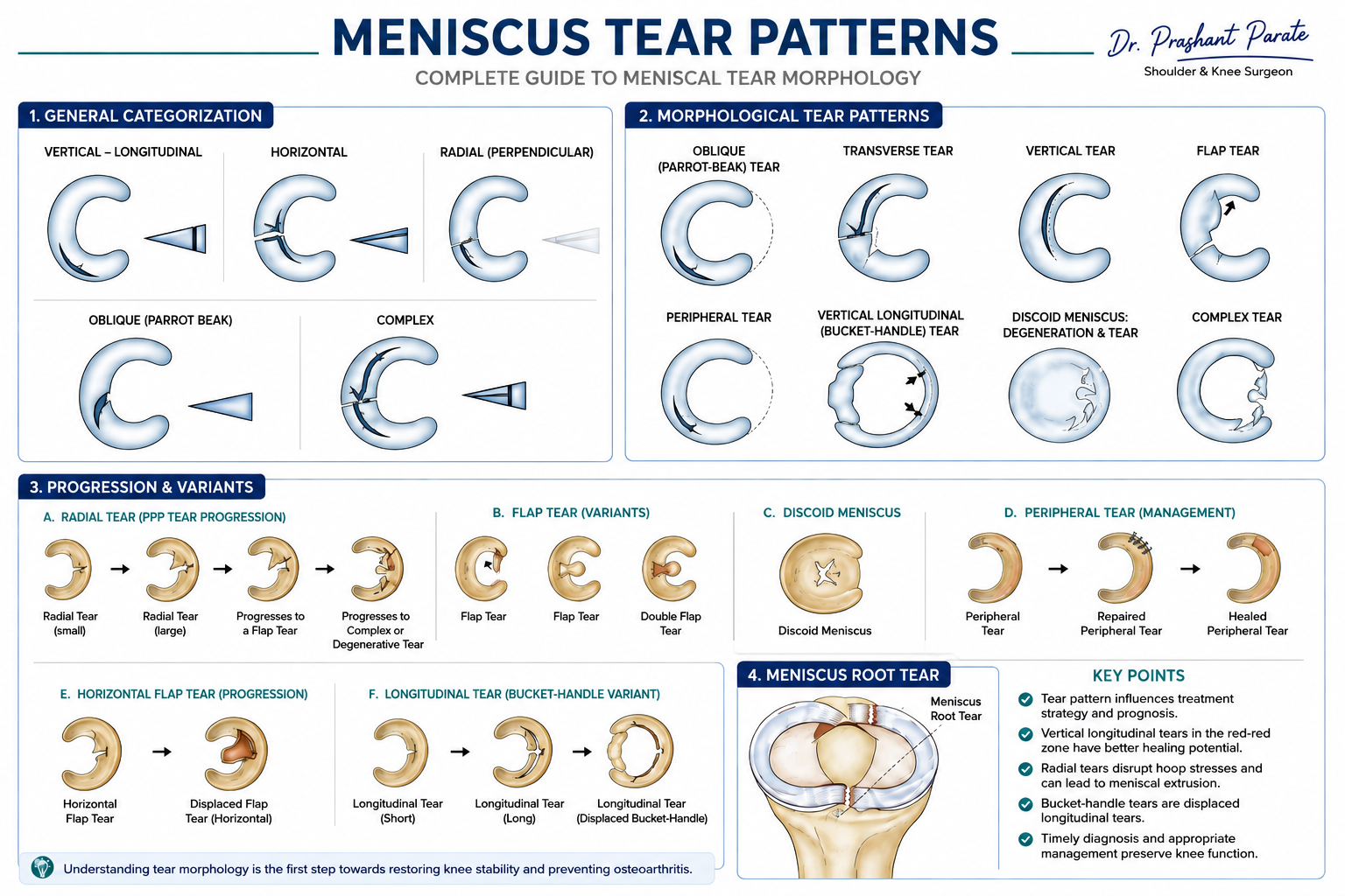
Longitudinal and bucket-handle tears run along the length of the meniscus and are the most reliably repairable. The tissue on both sides of the tear is usually healthy enough to hold sutures, and the blood supply is often adequate. A displaced bucket-handle tear needs to be reduced back into position first, and if the tissue quality is good, repair rates are high.
Peripheral vertical tears in the red zone are usually straightforward to repair and heal well.
Root tears, where the meniscus detaches from its anchor on the bone, are a special case. A root tear effectively turns a functioning meniscus into a non-functioning one, because without its attachment the meniscus cannot spread load. In an appropriately selected symptomatic patient without advanced arthritis, root repair should be strongly considered, because leaving it detached produces the same outcome as removing the entire meniscus.
Horizontal cleavage tears and partial-width radial tears are in the uncertain zone. They are worth attempting to repair, particularly in younger patients, even when the chance of healing is not high. The reasoning is the same: if it heals, you have saved years of cartilage life. If it does not, you trim it later, and you are no worse off than if you had trimmed it at the first operation.
Severely degenerative, fragmented and some complex or unstable flap tears may not provide enough healthy tissue for reliable fixation. Radial tears are different. Even complete radial tears can sometimes be repaired using modern techniques when tissue quality and the condition of the knee are favourable, because disruption of the circumferential fibres can severely impair the meniscus’s ability to transmit load. When repair is genuinely impossible, only the unstable non-viable portion is trimmed, preserving the maximum functional rim.
Repair techniques
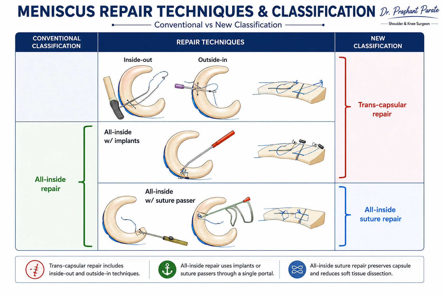
Three techniques exist, and the choice depends on where the tear is, how accessible it is, and the tissue quality.
All-inside repair
The suture device is passed entirely from within the joint through the arthroscope, avoiding the accessory incision required for traditional inside-out repair. All-inside repair is particularly useful for tears in the posterior part of the meniscus. I particularly favour it for many posterior horn tears, including selected lateral meniscus tears. Modern all-inside techniques can reduce the risk associated with posterior suture retrieval, although careful device depth and trajectory remain essential because important neurovascular structures still lie behind the knee. The implants are more expensive than conventional needles and sutures.
Inside-out repair
Long needles are passed through the meniscus from inside the joint and brought out through a small incision at the back of the knee. The sutures are tied on the capsule under direct vision. This has traditionally been the gold standard because it gives strong, precise suture placement. The disadvantage is the extra incision, and the need to carefully protect the nerve at the back of the knee during suture retrieval.
Outside-in repair
A needle is passed from outside the knee into the joint across the tear. This works best for tears at the front of the meniscus, where the other two techniques have difficulty reaching. It is used in selected cases only.
In practice, the surgeon may use more than one technique in the same knee, choosing the best approach for each part of the tear.
Ramp lesions: the tear that hides on MRI
A ramp lesion is a tear at the back of the medial meniscus where it attaches to the capsule. It is particularly associated with ACL injuries and can be subtle enough to miss on routine MRI.
During ACL reconstruction, the posterior compartment needs careful assessment because an unstable ramp lesion left unrepaired can compromise the result. These tears are usually repairable once identified, and repair at the time of ACL reconstruction is straightforward.
When repair fails: the role of alignment
A meniscus repair can fail for several reasons, and malalignment is one of the most important. If the leg is bowed (varus) or knock-kneed (valgus), the load is concentrated on one side of the joint, and that overloaded side is where the meniscus was repaired. The repair heals under load it cannot sustain, and it fails.
Significant malalignment may need to be corrected to protect the repair.
In selected patients with substantial malalignment, an osteotomy performed with or around the time of ligament and meniscus surgery can unload the repaired compartment and improve the mechanical environment for healing. Patients with associated cartilage damage can read more about knee arthritis and joint-preservation options. The amount of deformity, cartilage status, tear pattern, symptoms and patient age all influence this decision.
Other factors that reduce healing include poor tissue quality, tears that have been present for a long time, tears in the white-white zone, and inadequate rehabilitation. The combination of a chronic ACL injury with a degenerative meniscus tear in a malaligned knee is the scenario where repair is least likely to succeed, and where the decision between repair, partial removal and correction of alignment requires careful judgement.
The philosophy: repair first, excise later
Not every repair will heal, and that is accepted. The question is not whether the repair is guaranteed to succeed, but whether the patient is better off having attempted it.
Take a chance on repair even when the odds are uncertain.
If a repair heals, it adds years of cartilage protection. If it fails, the meniscus is trimmed at a later date, and the patient is no worse off than if it had been trimmed at the first operation. The only thing lost is time, and what is gained, if it works, is a functioning meniscus and a slower path to arthritis.
This philosophy applies most strongly in younger patients, where the consequences of early meniscectomy are measured in decades of accelerated wear. In an older patient with an already degenerative meniscus and established cartilage damage, a clean partial meniscectomy may be the more practical choice.
There is an important distinction between a meniscus that cannot be repaired and one that is difficult to repair. Modern meniscus surgery increasingly tries to distinguish between the two, because only the first justifies removal.
Root repair: a special priority
A meniscus root tear deserves particular mention because it is underdiagnosed and undertreated. The root is where the meniscus anchors to the bone, and without that anchor the meniscus extrudes outward and stops functioning as a load spreader. The biomechanical effect is the same as removing the entire meniscus.
When the cartilage is reasonably preserved and the patient is an appropriate candidate, anatomical root repair is generally preferred to leaving the meniscus functionally detached. The torn root is pulled back to its anatomical position and fixed to bone through a tunnel. This restores function and protects the cartilage. If the cartilage is already severely damaged, root repair may not reverse the process, and the treatment shifts towards managing the arthritis.
What happens when too much meniscus is lost
A young patient who has lost most of the medial or lateral meniscus may develop compartment-specific pain, reduced tolerance to loading, recurrent swelling and progressive cartilage overload. This is sometimes called post-meniscectomy syndrome, and it is a difficult long-term problem. This is one of the strongest reasons to preserve meniscal tissue at the original operation whenever it is reasonably possible.
Meniscus transplant: when nothing can be saved
In a young patient who has lost a large part of the medial meniscus, either from an irreparable tear or from a previous meniscectomy, and who now has pain on that side of the joint with intact cartilage, meniscus transplantation can be considered.
A donor meniscus, matched for size, is implanted into the knee and fixed to bone and capsule. This is not a routine procedure. It is reserved for patients who are young enough to benefit, have good alignment or are willing to have alignment corrected, have intact cartilage, and have a specific pattern of symptoms. The evidence supports its use as a bridge to delay arthritis and replacement, not as a permanent solution.
The final decision is made during arthroscopy
MRI is extremely useful, but it cannot always determine the final repair strategy. During arthroscopy I can directly assess the exact tear configuration, the tissue quality, whether the fragment can be reduced, the state of the capsular attachment, and any associated cartilage injury or additional tears that MRI did not show. For this reason, I discuss the possibilities with every patient before surgery: repair, partial removal, a combination of repair and limited trimming, or occasionally leaving a stable lesion alone. The final decision follows what the tear actually looks like, not what the MRI predicted.
How recovery differs when the meniscus is repaired
An ACL reconstruction alone allows relatively early weight bearing and range of motion. Adding a meniscus repair changes the rehabilitation significantly, because the repair needs protection while it heals.
| Aspect | ACL alone | ACL + meniscus repair* |
|---|---|---|
| Weight bearing | As tolerated from early on | Protected for 4 to 6 weeks |
| Bending limit | Progress as tolerated | Limited to 90 degrees for 4 to 6 weeks |
| Deep squatting | From around 3 months | Not before 3 months, often later |
| Return to sport | 9 to 12 months | May be delayed further |
*These restrictions are examples rather than a universal protocol. Some stable peripheral repairs may allow earlier weight bearing and motion, whereas radial, complex and especially root repairs may require substantially greater protection. The operative findings and repair type determine the final rehabilitation protocol.
The exact restrictions depend on the type of tear, the location of the repair, the technique used, and whether a root repair was performed. A root repair typically requires more protective early loading than a peripheral repair. The individual rehabilitation protocol issued after surgery specifies the exact limitations, and that protocol overrides any general guidance.
What you should ask your surgeon
If you have been told you need ACL reconstruction and have a meniscus tear as well, these questions are worth asking. You may also find this guide useful: how to choose the right orthopaedic surgeon for your shoulder or knee problem.
Is my meniscus tear repairable?
If it is not certain, will you attempt a repair before deciding to trim it?
What technique will you use, and why?
Is my leg alignment normal, or does it need correcting to protect the repair?
How will the repair change my recovery compared with ACL surgery alone?
If the meniscus cannot be saved, is transplant something to consider in the future?
Related Knee Guides
ACL Tear and ACL Reconstruction
Meniscus Tear: Repair or Removal?
ACL tear with a meniscus injury?
Whether the meniscus can be repaired depends on the tear pattern, blood supply, tissue quality and knee alignment. Bring your MRI.
Appointments by phone
Sainath Hospital, Moshi, PCMC
86000 05886
Mon to Fri, 10 am to 4 pm · Sat, 10 am to 12 noon
Jupiter Hospital, Baner, Pune
020 2799 2150
Mon to Sat, 5.30 pm to 6.30 pm · OPD 5
This guide is intended for patients and their families. It does not replace assessment and should not be used to make treatment decisions without a consultation. Last reviewed: August 2026 by Dr. Prashant Parate, DNB Orthopaedics, Fellowship in Arthroscopy & Sports Medicine (Thammasat University Hospital, Thailand). MMC Reg. 2006042129.